Clinical-Radiological Presentation 32

Question

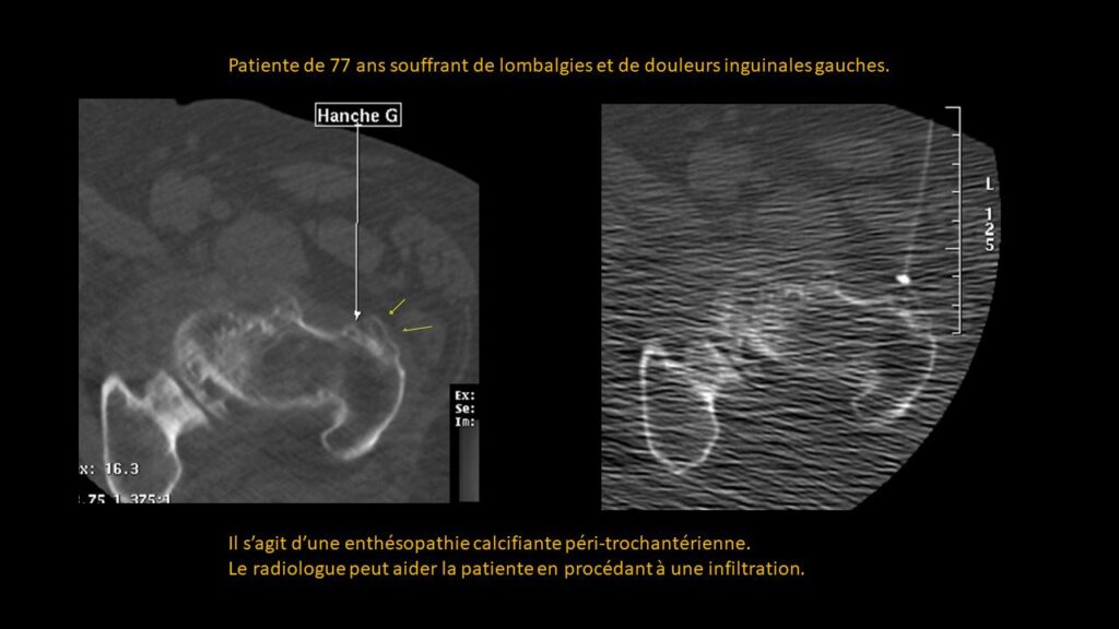
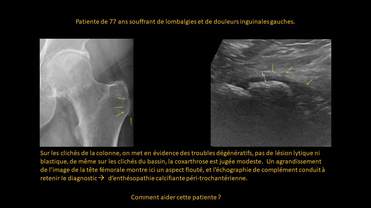
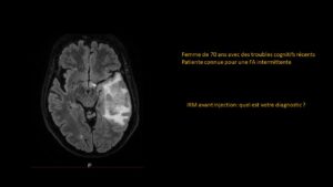
77-year-old patient experiencing lower back pain and left inguinal pain.

Imaging of the spine reveals degenerative changes, with no lytic or blastic lesions. Similarly, imaging of the pelvis shows mild coxarthrosis. A close-up view of the femoral head reveals a blurred appearance, and additional ultrasound imaging supports a diagnosis of peritrochanteric calcific enthesopathy.

ANSWER

This is a peritrochanteric calcific enthesopathy.
The radiologist can help the patient by performing an infiltration, specifically a corticosteroid injection.

Partagez :

Imagerie effectuée au CIL par le Dr Amir MEHDIZADE, spécialiste FMH en Radiologie

Spécialiste  en Neuroradiologie, Ancien Chef de Clinique aux Hôpitaux Universitaires de Genève  (HUG)
Médecin Consultant aux Hôpitaux  Universitaires de Genève  (HUG)
Clinical Fellow in Breast and Body Imaging, Memorial Sloan Kettering, NEW-YORK
Clinical Fellow in Musculoskelatal Imaging, Hospital for Special Surgery, Cornel University, NEW-YORK

UNITES FONCTIONNELLES

RADIOLOGIE CONVENTIONNELLE
IRM CORPS ENTIER
SCANNER LOW DOSE
MAMMOGRAPHIE MICRODOSE
ECHOGRAPHIE-DOPPLER
FIBROSCAN
PONCTIONS INFILTRATIONS
DENSITOMETRIE

Les Pathologies à travers l'Objectif Radiologique