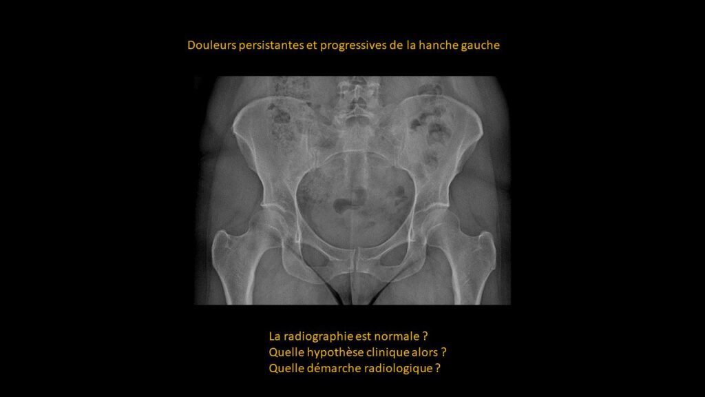
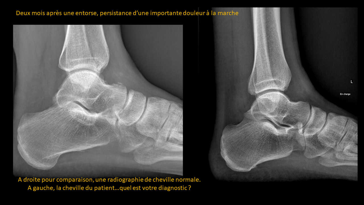
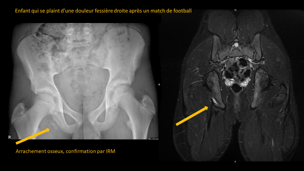
Clinical-Radiological Presentation 18

Question

Persistent and progressive pain in the left hip

Is the X-ray normal?
What is the clinical hypothesis then?
What radiological approach should be taken?
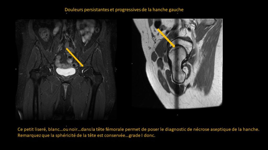
ANSWER

Look at the MRI: This small line, white or black, in the femoral head allows the diagnosis of avascular necrosis of the hip.
Note that the sphericity of the head is preserved, so it is a Grade I.

Partagez :

Imagerie effectuée au CIL par le Dr Amir MEHDIZADE, spécialiste FMH en Radiologie

Spécialiste  en Neuroradiologie, Ancien Chef de Clinique aux Hôpitaux Universitaires de Genève  (HUG)
Médecin Consultant aux Hôpitaux  Universitaires de Genève  (HUG)
Clinical Fellow in Breast and Body Imaging, Memorial Sloan Kettering, NEW-YORK
Clinical Fellow in Musculoskelatal Imaging, Hospital for Special Surgery, Cornel University, NEW-YORK

UNITES FONCTIONNELLES

RADIOLOGIE CONVENTIONNELLE
IRM CORPS ENTIER
SCANNER LOW DOSE
MAMMOGRAPHIE MICRODOSE
ECHOGRAPHIE-DOPPLER
FIBROSCAN
PONCTIONS INFILTRATIONS
DENSITOMETRIE

Les Pathologies à travers l'Objectif Radiologique