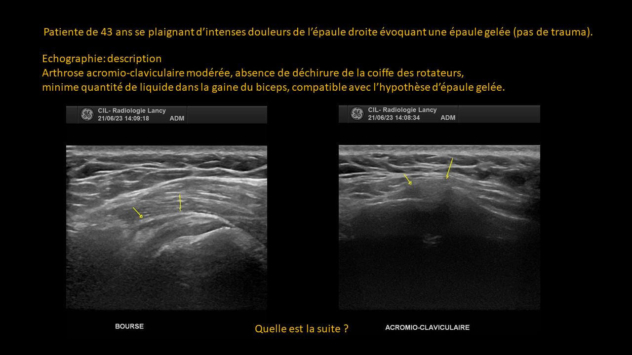
Clinical-Radiological Presentation 10

Question

Dizziness and Headaches Lead to an MRI Request

For comparison, on the right is a normal image. And on the left, follow the arrows…

What is this part of the brain called and what do you see there?
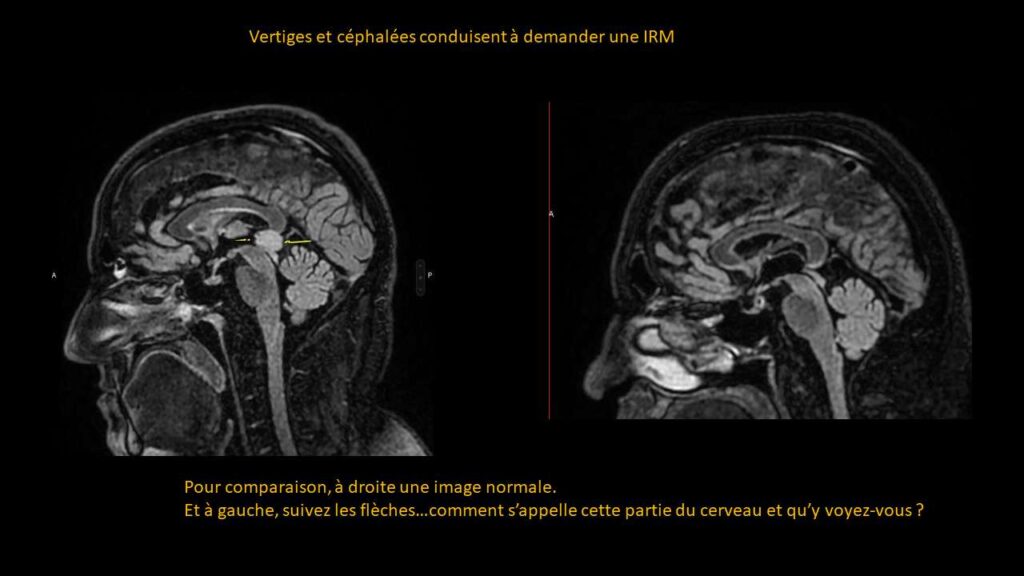
ANSWER

It is the pineal gland, which is cystic.
There is no compression of the cerebrospinal fluid (CSF) pathways, no upstream dilation, and no hydrocephalus.

This finding does not really explain the symptoms… it is more of an incidental finding!

Partagez :

Imagerie effectuée au CIL par le Dr Amir MEHDIZADE, spécialiste FMH en Radiologie

Spécialiste  en Neuroradiologie, Ancien Chef de Clinique aux Hôpitaux Universitaires de Genève  (HUG)
Médecin Consultant aux Hôpitaux  Universitaires de Genève  (HUG)
Clinical Fellow in Breast and Body Imaging, Memorial Sloan Kettering, NEW-YORK
Clinical Fellow in Musculoskelatal Imaging, Hospital for Special Surgery, Cornel University, NEW-YORK

UNITES FONCTIONNELLES

RADIOLOGIE CONVENTIONNELLE
IRM CORPS ENTIER
SCANNER LOW DOSE
MAMMOGRAPHIE MICRODOSE
ECHOGRAPHIE-DOPPLER
FIBROSCAN
PONCTIONS INFILTRATIONS
DENSITOMETRIE

Les Pathologies à travers l'Objectif Radiologique