Clinical-Radiological Presentation 01

Question

Two months after an ankle sprain, the patient continues to experience significant pain while walking.
On the right, we have a normal ankle X-ray for comparison.
On the left, we have the patient’s ankle X-ray… what is your diagnosis?
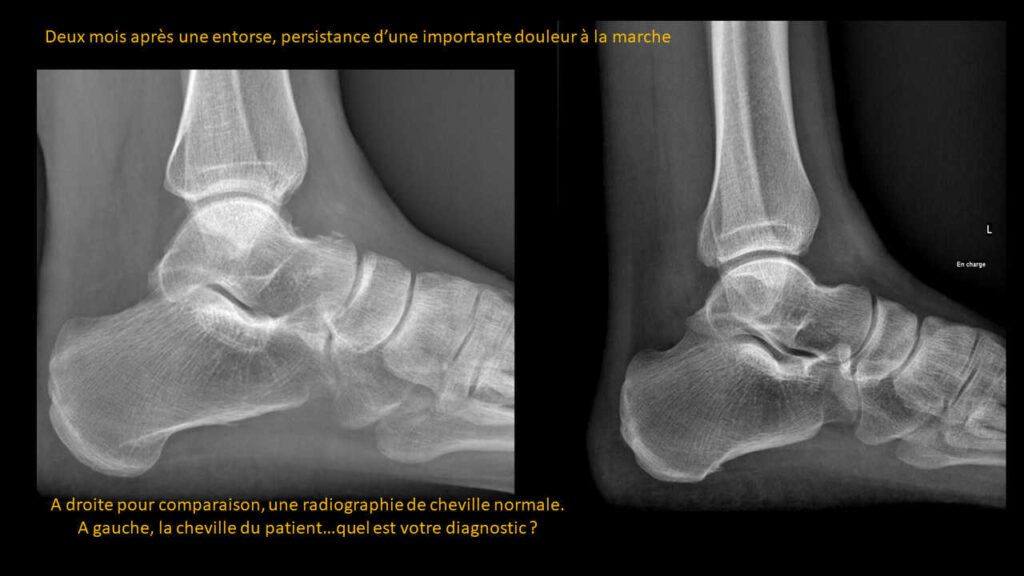
ANSWER

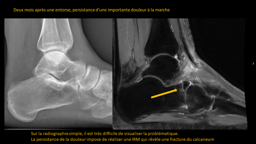
On the plain X-ray, it is very difficult to visualize the issue.

The persistence of pain necessitates an MRI, which then reveals a calcaneal fracture.

Partagez :

Imagerie effectuée au CIL par le Dr Amir MEHDIZADE, spécialiste FMH en Radiologie

Spécialiste  en Neuroradiologie, Ancien Chef de Clinique aux Hôpitaux Universitaires de Genève  (HUG)
Médecin Consultant aux Hôpitaux  Universitaires de Genève  (HUG)
Clinical Fellow in Breast and Body Imaging, Memorial Sloan Kettering, NEW-YORK
Clinical Fellow in Musculoskelatal Imaging, Hospital for Special Surgery, Cornel University, NEW-YORK

UNITES FONCTIONNELLES

RADIOLOGIE CONVENTIONNELLE
IRM CORPS ENTIER
SCANNER LOW DOSE
MAMMOGRAPHIE MICRODOSE
ECHOGRAPHIE-DOPPLER
FIBROSCAN
PONCTIONS INFILTRATIONS
DENSITOMETRIE

Les Pathologies à travers l'Objectif Radiologique