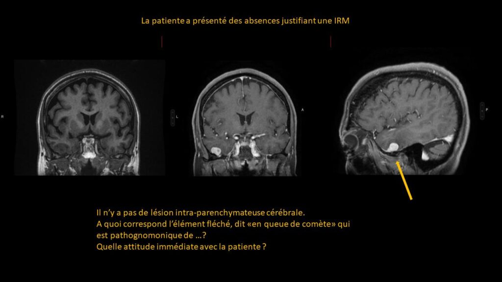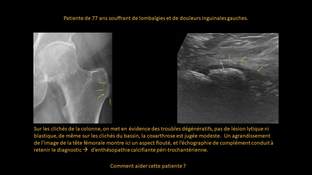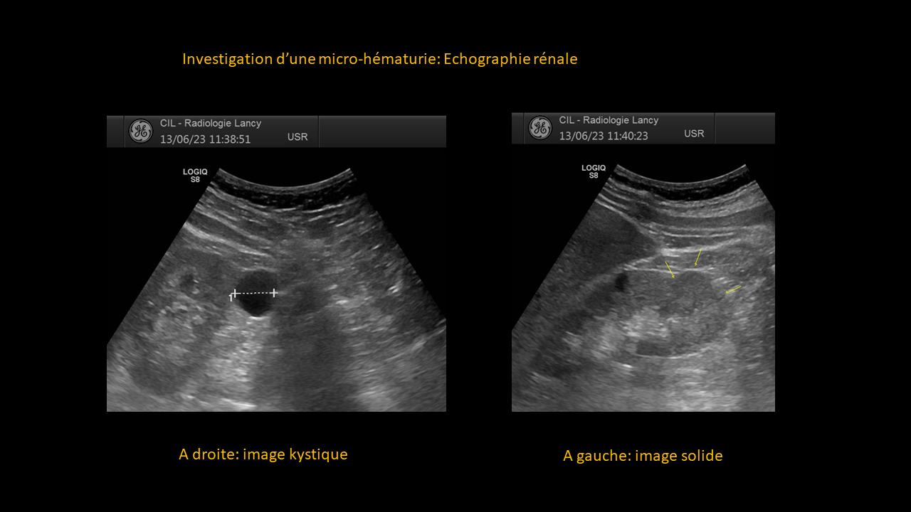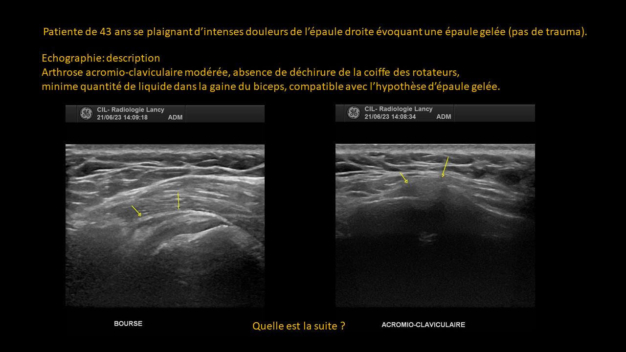
Question
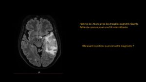
The patient experienced episodes of absence justifying an MRI.
There is no intra-parenchymal cerebral lesion.
What does the arrowed element, referred to as a “comet tail,” which is pathognomonic of… correspond to?
What immediate approach should be taken with the patient?
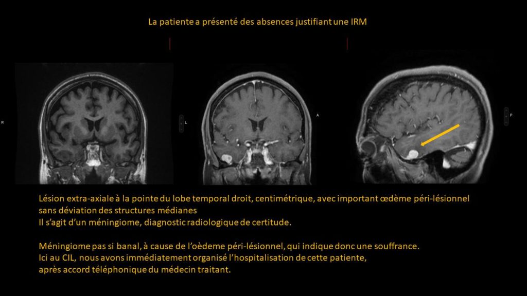
ANSWER
Extra-axial lesion at the tip of the right temporal lobe, measuring a few centimeters, with significant perilesional edema without deviation of the midline structures. It is a meningioma, identified on the MRI.
This meningioma is not so trivial due to the perilesional edema, which indicates suffering. Here at the CIL, we immediately organized the hospitalization of this patient after obtaining telephone consent from her primary care physician.