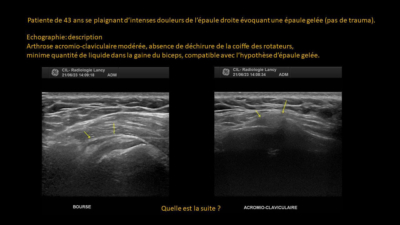
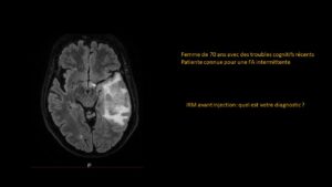
Clinical-Radiological Presentation 09

Question

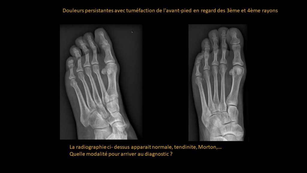
Persistent pain with swelling of the forefoot in the area of the 3rd and 4th rays. No trauma.

The X-ray below appears normal. Possibilities include tendinitis, Morton’s neuroma, etc.

Which imaging modality should be used to reach the diagnosis?

ANSWER

MRI: Proximal diaphyseal-metaphyseal junction of the 4th ray: No fracture line, no arthritis, and no soft tissue infiltration.

Metatarsal hallux: Focal sub-capital hyperpressure with new bursopathy.

CONCLUSION = Overall appearance suggestive of static disorders.

Partagez :

Imagerie effectuée au CIL par le Dr Amir MEHDIZADE, spécialiste FMH en Radiologie

Spécialiste  en Neuroradiologie, Ancien Chef de Clinique aux Hôpitaux Universitaires de Genève  (HUG)
Médecin Consultant aux Hôpitaux  Universitaires de Genève  (HUG)
Clinical Fellow in Breast and Body Imaging, Memorial Sloan Kettering, NEW-YORK
Clinical Fellow in Musculoskelatal Imaging, Hospital for Special Surgery, Cornel University, NEW-YORK

UNITES FONCTIONNELLES

RADIOLOGIE CONVENTIONNELLE
IRM CORPS ENTIER
SCANNER LOW DOSE
MAMMOGRAPHIE MICRODOSE
ECHOGRAPHIE-DOPPLER
FIBROSCAN
PONCTIONS INFILTRATIONS
DENSITOMETRIE

Les Pathologies à travers l'Objectif Radiologique